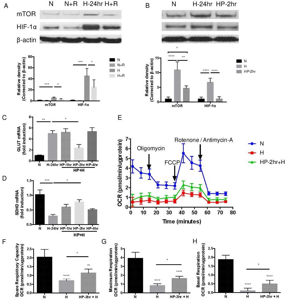
Fig. 6. Hypoxic preconditioning inhibits HIF-1α activation by modulating mTOR and decreasing glycolysis in MIO-M1 cells. (A) Exposure of MIO-M1 cells to 1% O2 hypoxia 24 hours (H-24hr), the increased expression of mTOR in accordance with HIF-1α compare to normoxia (N) and both (Normoxia with Rapamycin: N+R; Hypoxia with Rapamycin: H+R) were inhibited through pretreatment with rapamycin (100 nM) at protein level (n=3 per group). (B) HP-2 hours (HP-2hr) could inhibit the expression of HIF-1α through suppressing mTOR expression, decreased glycolysis and improves mitochondrial function in comparison with hypoxic treatment (n=3 per group). (C, D) Exposure MIO-M1 cells to hypoxia increased gene expression of GLUT and SDHD compared to normoxic treatment. However, HP-2 hours partially reversed the expression in comparison with hypoxia (n=4 per group). (E) Representative mitochondrial stress test measured with sequential injections of oligomycin, FCCP and rotenone/antimycin A. (F) Spare-respiratory capacity was calculated as the difference between maximal and basal OCR. (G) Maximal respiration was calculated as difference between maximal OCR value post-injection of FCCP and non-mitochondrial OCR. (H) Basal respiration was calculated as the difference between first OCR measurement and non-mitochondrial OCR. Data represents means ± SD of relative values vs control from 3 independent experiments. P<0.05; **P<0.01; ***P<0.001; ****P<0.0001, statistical analysis was performed with one-way ANOVA with Dunn's test for multiple comparisons.